Gallery

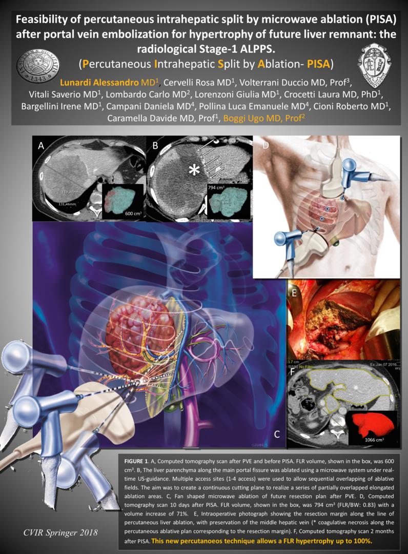
SPLIT EPATICO PERCUTANEO - P.I.S.A.

 Articolo in Stampa su Cardiovascular and Interventional Radiology (CVIR 2018)

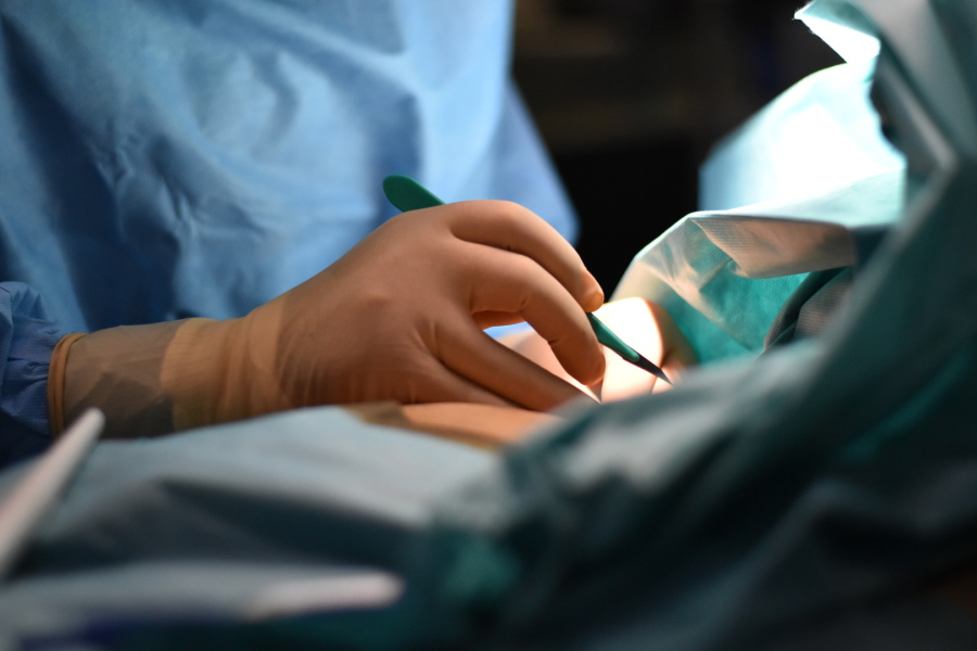
TECNICA DI SPLIT EPATICO PERCUTANEO ECOGUIDATO

"PISA"

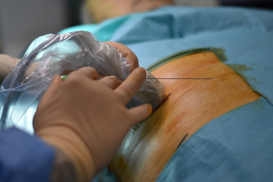
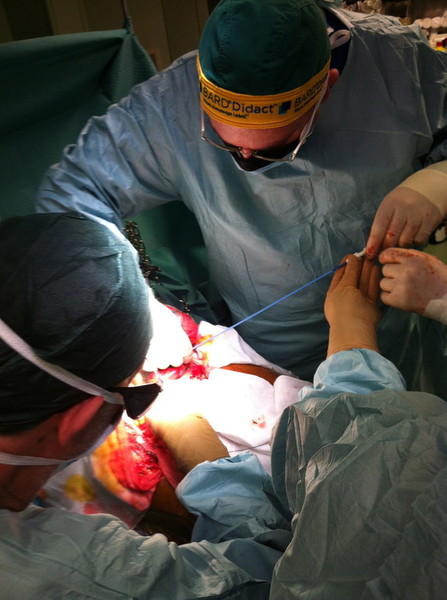
1 settimana dopo l'embolizzazione portale il fegato viene diviso seguendo il futuro piano chirurgico con un taglio ablativo percutaneo mediante microonde. Il paziente deambula

dopo poche ore dall'intervento che viene effettuato in sedazione profonda.

Il fegato sano raggiungerà volumi superiori al 100% del volume iniziale (63-110%) permettendo al chirurgo di effettuare l'epatectomia maggiore con un minor rischio

di insufficienza epatica post-operatoria.

Questa tecnica innovativa è stata ideata

dal Dott. Alessandro Lunardi assieme al Prof. Ugo Boggi nel 2015. 


CVIR 2018 Springer: http://rdcu.be/Fmot

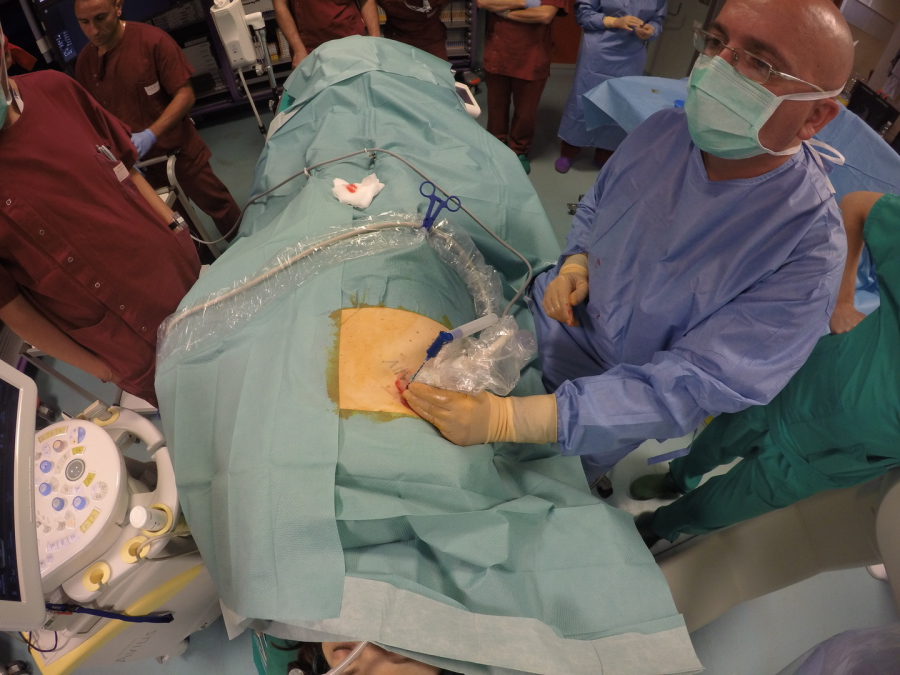
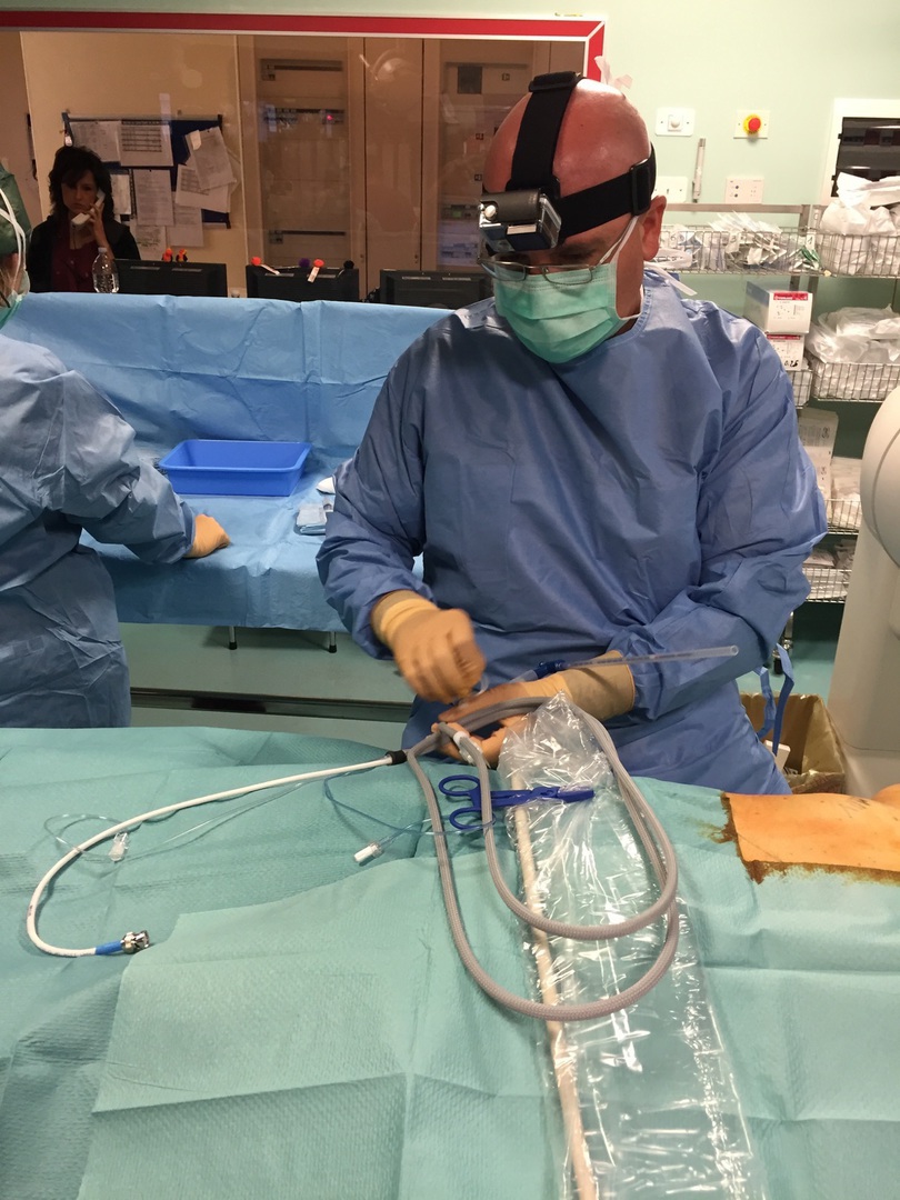
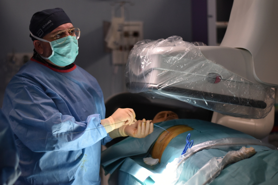
Primo intervento di "P.I.S.A." effettuato dal Dott. Lunardi nel 2015

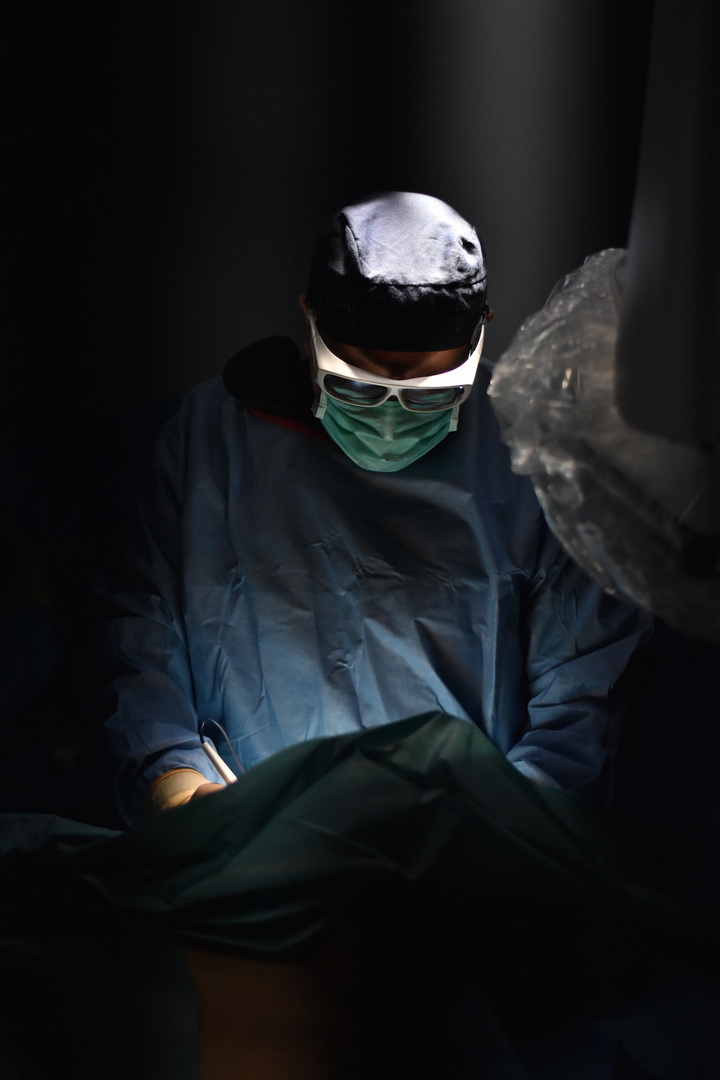
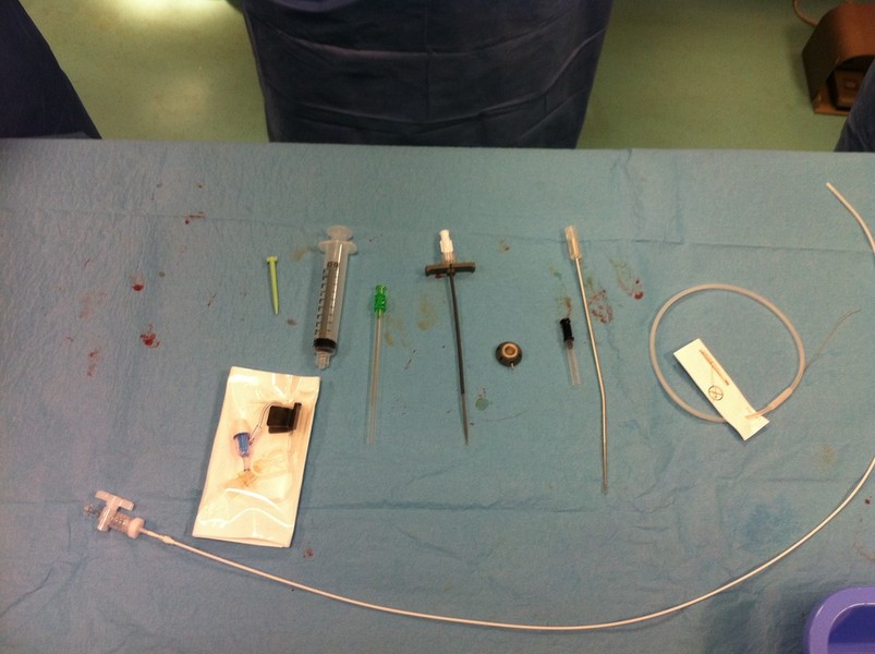
INTERVENTISTICA BILIARE PERCUTANEA

TECNICA MININVASIVA

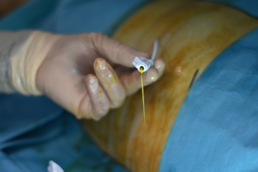
Puntura ecoguidata della Via Biliare - Radiologia Interventistica

Interventistica Biliare Pecutanea - Radiologia Interventistica

Accessi mininvasivi - Radiologia Interventistica

Drenaggio Biliare - Radiologia Interventistica

This female patient underwent biliary and papillary reconstruction in 1990 for lithiasis of the common bile duct.  After 26 years she develop papillary and  lower third common bile duct benign stricture with clinically relevant cholangitis and cholestasis.   The patient have been reffered to our Interventional Radiology Unit for PTC and bilioplasty. Restenosis occurred soon after bilioplasty.  Then I deployed a percutaneous transpapillary polydioxanone biodegradable stent 10x60mm.
Stent will degrade during next 3 months and then progressively disappear . 


n This is the 10th benign biliary stricture I successfully treat with this new promisingdevice during 2016.


The Zilver Vena Venous Self-Expanding Stent is the world’s first-ever stent designed to specifically treat symptomatic iliofemoral venous outflow obstruction. I used this device in a off label setting for this hemodyalisis patient with rapid recurrent strong subclavian vein restenosis after angioplasty. I hope to take the advantage of this features to solve problem of my patient:
large stent diameter to fit this big vessel (14mm)
consistently strong radial force from end to end
flexibility and kink-resistance

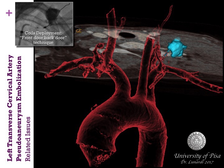
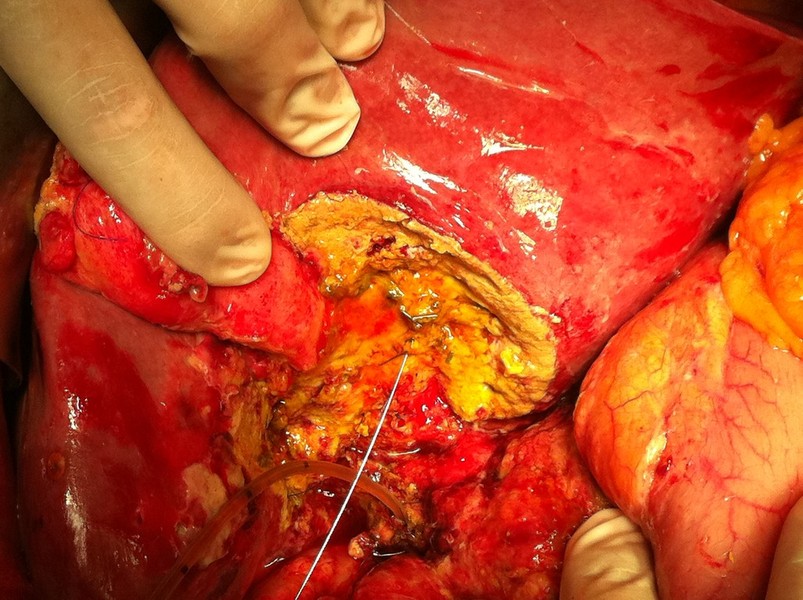
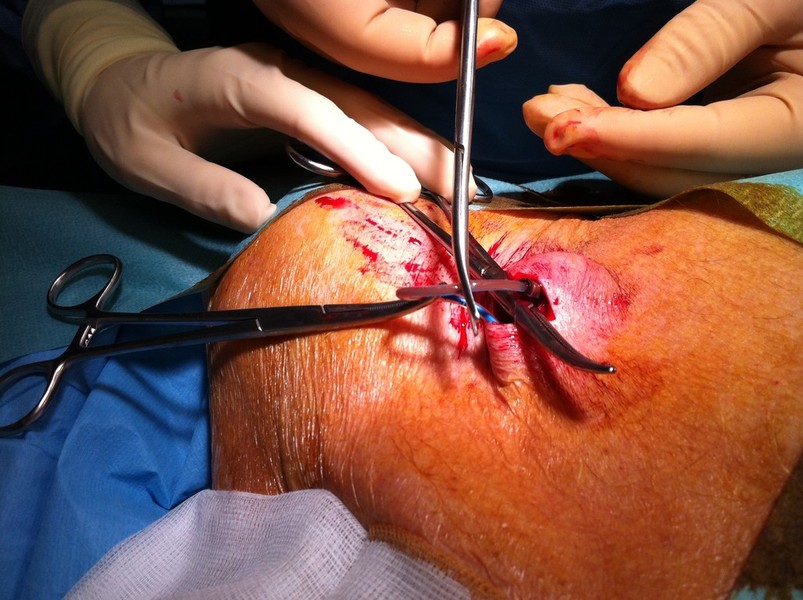
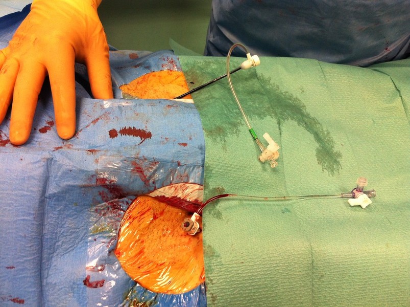
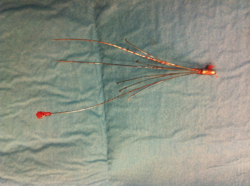
This young female patient developed a left transverse cervical artery pseudoaneurysm after the unsuccessful attempt of ipsilateral Internal Jugular vein catheterization during the hospital stay for orthotopic liver transplantation.

Main related Issues to this kind of embolization are:
Instability of catheters and  narrow curves at the origin of the target artery
Potential risk of brain embolization related to proximity of vertebral artery origin (i,.e. blood clots, gas bubbles or migration of liquid embolic agents)
Possible solutions are:
Use of long sheath vascular introducers (for example 6Fx90cm)
Continuous heparinized drip infusion of vascular introducer
Avoid the use of liquid embolic agents like acrylic glue mixtures or GelFoam
The Final solution seem to be Stand-alone coils embolization!


PHOTOS GALLERY


CVIR

Journal of Cardiovascular and Interventional Radiology

RADIOLOGY 

Stampa | Mappa del sito
© RadiologiaInterventisticaOnline - Dott. Alessandro Lunardi Radiologo Interventista Dirigente Medico 1° Livello U.O. Radiologia Interventistica - Azienda Ospedaliero Universitaria Pisana (AOUP) Pisa